
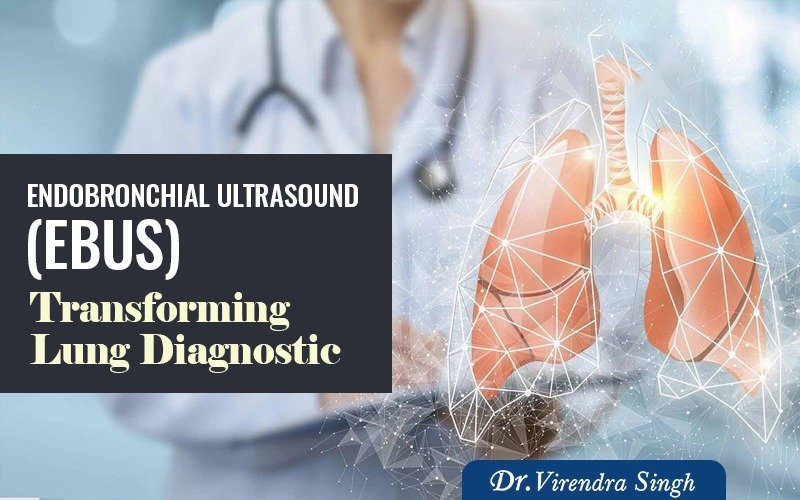
Endobronchial Ultrasound (EBUS) is a minimally invasive but highly effective procedure to diagnose lung cancer, illnesses, and infections that cause the chest’s lymph nodes (glands) to expand. The Department of Interventional Pulmonology at Asthma Bhawan is one of the first centers in North India to offer EBUS. We take it upon ourselves to provide some information about this cutting-edge diagnostic tool that is assisting professionals worldwide. By combining bronchoscopy with real-time ultrasound imaging, EBUS allows physicians to visualize the airways, lungs, and surrounding lymph nodes with exceptional clarity. This innovative technique minimizes patient discomfort, recovery time, and related dangers by enabling accurate needle biopsies without the need for surgical intervention.
EBUS has become a game-changer in early diagnosis and treatment planning, enhancing outcomes through safer, faster, and more targeted lung disease management. According to Asthma Bhawan, EBUS is a minimally invasive procedure that combines bronchoscopy with real-time ultrasound to examine the lungs and surrounding lymph nodes with precision. Infections, lung cancer, and unexplained lung disorders can all be detected and staged earlier because of EBUS, which makes precise needle biopsies possible.
What are the Types of Endobronchial Ultrasound?
There are two primary types of endobronchial ultrasound, each with special traits and uses:
-
Radial Probe EBUS (RP-EBUS)
A specialized bronchoscope with a radial ultrasonic probe at the distal end is used for RP-EBUS. This form of EBUS provides a 360-degree view of the airway walls and adjacent structures, allowing for thorough imaging and evaluation. RP-EBUS is very helpful for evaluating peripheral lung lesions, identifying bronchial wall anomalies, and directing diagnostic techniques such as needle aspiration or biopsy.
-
Convex Probe EBUS (CP-EBUS)
CP-EBUS utilizes a bronchoscope with a convex ultrasound probe located at the tip of the bronchoscope. A forward-viewing ultrasound image is provided by CP-EBUS, in contrast to RP-EBUS, which makes it possible to see structures right in front of the bronchoscope. CP-EBUS is frequently used to assess mediastinal lymph nodes, identify anomalies in the mediastinum, and direct diagnostic techniques such as needle aspiration or biopsy.
Why is EBUS Used?
EBUS is commonly used to diagnose and stage lung cancer, assess mediastinal lymph nodes for metastasis, and evaluate other conditions affecting the airways and surrounding tissues. It enables doctors to take fluid or tissue samples from the lungs and nearby lymph nodes without the need for traditional surgery. The samples can be used for:
- Diagnosing infections such as tuberculosis
- Diagnosing and staging lung cancer
- Diagnosing other cancers, such as lymphoma
- Diagnosing inflammatory diseases such as sarcoidosis
What are the Benefits of an Endobronchial Ultrasound?
When it comes to diagnosing and treating lung and mediastinal disorders, endobronchial ultrasound has many advantages. The key benefits of EBUS include:
- Minimally Invasive: EBUS is a minimally invasive method that eliminates the need for more invasive surgical procedures by employing a flexible bronchoscope that is inserted through the mouth or nose.
- Real-Time Imaging: Healthcare professionals can see anomalies like tumors, lymph nodes, or lesions with excellent resolution and precision due to EBUS’s real-time ultrasound imaging of the airways and surrounding tissues.
- Precise Targeting: EBUS enables accurate targeting of lesions or lymph nodes within the airways or mediastinum, facilitating staging, accurate diagnosis, and guidance of therapeutic interventions.
- Comprehensive Evaluation: Lung cancer, infections, and other respiratory disorders can be diagnosed and treated with the use of EBUS, which enables a thorough assessment of mediastinal lymph nodes as well as central and peripheral pulmonary abnormalities.
- Reduced Need for Surgical Biopsy: EBUS can often provide sufficient diagnostic information without the need for more invasive surgical procedures, such as thoracoscopy or mediastinoscopy, reducing recovery time, patient discomfort, and healthcare costs.
- Quick Results: Procedures guided by EBUS, including needle aspiration or biopsy, can yield quick findings, enabling prompt diagnosis and treatment planning—especially crucial in suspected cancer patients.
- Cost-Effective: Compared to other diagnostic procedures, EBUS is very cost-effective and also has minimal complications.
What to Expect?
Before the Procedure
- Medical History Review: The patient’s medical history, including any allergies or underlying medical disorders, will be reviewed by the doctor, who will also talk about any worries the patient may have regarding the surgery.
- Consent: The patient will be asked to sign a consent form, indicating that they understand the procedure, its risks, and benefits.
- Preparation: The doctor will provide instructions on how to prepare for the EBUS procedure. This can entail halting some drugs, including blood thinners, for a while and fasting for a predetermined amount of time.
- Anaesthesia: Usually, sedation or general anesthesia is used during EBUS to provide patient comfort and relaxation.
After the Procedure
- Recovery: Following the procedure, you will be brought to a recovery area where you will be closely watched while the anesthesia or sedative wears off.
- Observation: Your healthcare provider will monitor you for any instant complications or side effects, such as difficulty breathing, bleeding, or low blood pressure.
- Discharge Instructions: You will receive post-procedure care instructions, including any dietary or drinking restrictions, medication schedules, and follow-up visits, after you are completely awake and stable.
- Follow-up: A follow-up visit could be planned for you to go over the procedure’s outcomes and talk about any additional treatment or management strategies that might be required.
FAQs
What is EBUS?
A minimally invasive procedure using ultrasound with a bronchoscope to examine and biopsy lung tissues and lymph nodes.
Why is EBUS done?
To diagnose lung cancer, infections, or enlarged lymph nodes and guide treatment decisions.
How is EBUS different from bronchoscopy?
EBUS uses ultrasound to see beyond the airway walls and take accurate tissue samples.
Is EBUS safe?
Yes, it’s generally safe with rare complications like minor infection or bleeding.
How long does it take?
Usually 30-60 minutes, with a short recovery time.